
Gray or silver scale nail pitting 70% of affected children have family history of psoriasis 2 Tinea corporis (annular lesions with well-defined, scaly, often reddish margins commonly pruritic) Differential Diagnosis of Tinea Infections Differential diagnosis
3. TINEA CAPITIS SCALP SKIN
2 Skin biopsy with periodic acid–Schiff (PAS) stain may rarely be indicated for atypical or persistent lesions. Cultures are usually not necessary to diagnose tinea corporis. Conversely, if a nonfungal lesion is treated with an antifungal cream, the lesion will likely not improve or will worsen. Worsening after empiric treatment with a topical steroid should raise the suspicion of a dermatophyte infection. 2, 3 A potassium hydroxide (KOH) preparation is often helpful when the diagnosis is uncertain based on history and visual inspection. Tinea corporis may be mistaken for many other skin disorders, especially eczema, psoriasis, and seborrheic dermatitis ( Table 2). Lesions may be single or multiple and the size generally ranges from 1 to 5 cm, but larger lesions and confluence of lesions can also occur.
3. TINEA CAPITIS SCALP PATCH
Tinea corporis (ringworm) typically presents as a red, annular, scaly, pruritic patch with central clearing and an active border ( Figure 1). Tinea Corporis, Tinea Cruris, and Tinea Pedis.In one survey, tinea was the skin condition most likely to be misdiagnosed by primary care physicians. Tinea infections can be difficult to diagnose and treat. The most common infections in prepubertal children are tinea corporis and tinea capitis, whereas adolescents and adults are more likely to develop tinea cruris, tinea pedis, and tinea unguium (onychomycosis). Uncommon fungal skin infections that involve other organs (e.g., blastomycosis, sporotrichosis) Pityriasis versicolor (formerly tinea versicolor) caused by Malassezia species Tinea incognito (altered appearance of dermatophyte infection caused by topical steroids)Ĭandida (yeast) and mold, which may cause onychomycosis or coexist in a dystrophic nail Tinea barbae (beard infection in male adolescents and adults) Tinea manuum (commonly presents with “one-hand, two-feet” involvement) Tinea corporis (ringworm), includes tinea gladiatorum and tinea faciei Dermatophytes include three genera: Trichophyton, Microsporum, and Epidermophyton. Dermatophytes are usually limited to involvement of hair, nails, and stratum corneum, which are inhospitable to other infectious agents. Tinea unguium is more commonly known as onychomycosis. Tinea versicolor (now called pityriasis versicolor) is not caused by dermatophytes but rather by yeasts of the genus Malassezia.

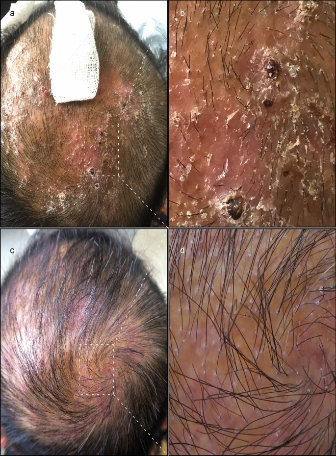
Tinea is usually followed by a Latin term that designates the involved site, such as tinea corporis and tinea pedis ( Table 1). The term tinea means fungal infection, whereas dermatophyte refers to the fungal organisms that cause tinea. Failure to treat kerion promptly can lead to scarring and permanent hair loss. However, kerion should be treated with griseofulvin unless Trichophyton has been documented as the pathogen. Oral terbinafine is first-line therapy for tinea capitis and onychomycosis because of its tolerability, high cure rate, and low cost. Tinea corporis, tinea cruris, and tinea pedis generally respond to inexpensive topical agents such as terbinafine cream or butenafine cream, but oral antifungal agents may be indicated for extensive disease, failed topical treatment, immunocompromised patients, or severe moccasin-type tinea pedis. Physicians should confirm suspected onychomycosis and tinea capitis with a potassium hydroxide preparation or culture. For example, tinea corporis can be confused with eczema, tinea capitis can be confused with alopecia areata, and onychomycosis can be confused with dystrophic toe-nails from repeated low-level trauma. The clinical diagnosis can be unreliable because tinea infections have many mimics, which can manifest identical lesions.


Tinea infections are caused by dermatophytes and are classified by the involved site.


 0 kommentar(er)
